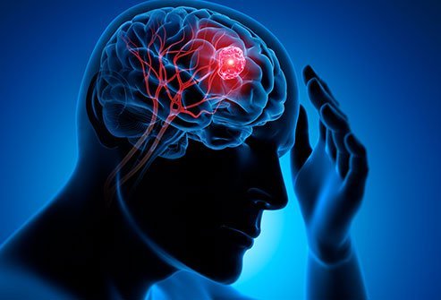
TYPES OF BRAIN SURGERY
Neurosurgery or Brain surgery is a critical and complicated medical procedure performed by trained neurosurgeons in a highly specialized environment. This involves repairing structural problems in the brain to correct any abnormalities such as tumors or aneurysms.
Brain surgery is performed in certain critical conditions such as:
- To prevent bleeding inside the brain due to aneurysm
- Removal of brain tumor
- Treat a pinched nerve in the brain area
- Drain an abscess or blood due to an injury
- Remove pressure on the brain due to blood or fluid accumulation
CONDITIONS REQUIRING BRAIN SURGERY
Structural abnormalities in the brain require brain surgery as they would impact organs being controlled by the affected part of the brain. Certain critical conditions that would require brain operation are:
- Congenital brain defects – Malformation of blood vessel at the connections between veins and arteries is formed in the brain region
- Internal bleeding in the brain from an aneurysm causing an arterial wall to weakens and ruptures
- Blood clots and bleeding in the brain –Brain operation helps to prevent auxiliary trauma to brain tissue surrounding the affected area
- Epidural or subdural abnormalities – Including hematomas
- Edema of the brain – It is the buildup of fluid around the brain leading to swelling and recurring headaches and pressure to the brain
- Brain tumors – These are removed through critical brain surgery, mainly in the case of cancer formations
- Rare cases of epilepsy – This is caused by an abnormality in the brain structure, or pressure on a nerve
- Neuropathic pain – This would occur when a nerve between the brain and the spine is damaged from a stroke, tumor or accident.
- Infection in surrounding areas or brain tissue – Caused the area in the brain to be filled with the infected fluid.
- Parkinson’s disease – Leads to the nervous signals essential to control motor function are excessively weak or abnormal.
Not all the above conditions essentially require brain surgery but it is proven that brain surgery has helped in minimizing the risk of more serious health problems.
TYPES OF BRAIN SURGERY
The brain surgery would be depending on the part of the brain and the condition being treated by the neurosurgeon.
1. Minimally Invasive Brain Surgery
This is also referred to as Keyhole surgery which is based on the concept of carefully removing brain & skull base tumors through smaller yet more precise openings.
This would help in minimizing the damage to the surrounding scalp, brain, blood vessels, and nerves. This procedure provides better results in comparison to normal open surgical procedures.
Minimally invasive neuro-navigation technology helps in reducing incisions as well as allows the surgeons to cosmetically hide scars. Some of the common minimally invasive surgical techniques being used by neurosurgeons are:
Endoscopic endonasal surgery – The technique involves a thin, flexible tube that has a light source along with a camera known as endoscope. These are threaded through the nose and sinus to enable the surgeon to access regions of the brain which are hard to reach using traditional surgical approaches requiring incisions mainly at the base of the skull or top of the spine. The endoscope would illuminate the regions of the brain with the help of specifically designed tools inserted through the nose to remove tumors or lesions.
Craniotomy – This is the surgical removal of a section of the bone (known as bone flap) from the skull using a specialized tool which exposes the brain. The bone flap is only removed temporarily which is replaced again after surgery. This procedure requires the direction from computers and imaging devices such as magnetic resonance imaging (MRI) or computerized tomography (CT) scans, to reach the affected location within the brain with the help of a frame placed onto the skull or a frameless system (using superficially placed markers or landmarks on the scalp). When these imaging procedures are used along with the craniotomy procedure is referred to as stereotactic craniotomy. Craniotomy is useful in removal of tumors, clip off an aneurysm, drain blood or fluid accumulated due to an infection as well as remove abnormal brain tissue.
Craniotomy is of various types such as:
- Extended Bifrontal Craniotomy
- Minimally Invasive Supra-Orbital “Eyebrow” Craniotomy
- Retro-Sigmoid “Keyhole” Craniotomy Orbitozygomatic Craniotomy
- Translabyrinthine Craniotomy
Deep Brain Stimulation – This technique involves implanting electrodes within affected areas of your brain. These electrodes produce electrical impulses responsible for regulating abnormal impulses affecting some cells and chemicals within the brain. The amount of stimulation produced during the procedure is controlled by a small device implanted under the skin in your upper chest with the help of a wire connecting the device to the electrodes in your brain. Deep Brain Stimulation is helpful in lowering the symptoms of tremor, slowness, stiffness, and walking issues resulting from Parkinson’s disease, dystonia, or essential tremor. Successful Deep brain stimulation helps people to possibly lower their medications as well as improve their quality of life. Other critical conditions like Dystonia, Epilepsy, Essential tremor and Obsessive-compulsive disorder are treated using this technique.
A deep brain stimulation technique involves 3 main parts which are implanted inside your body:
- Neuro-stimulator – This is a programmable battery-powered device implanted under the skin of your chest below the collarbone or in the abdomen. This generates electric pulses.
- Lead – This is a wire coated with a number of electrodes at its tip, implanted inside the brain. This joins to an extension wire through a tiny hole in the skull which delivers electric pulses to the brain tissue.
- Extension – This is an insulated wire implanted underneath the skin and runs from the scalp, behind the ear, down the neck, and to the chest. This would join the lead to the neuro-stimulator.
Neuroendoscopy – The neurosurgeon uses this technique to remove the tumor with the help of an endoscope (a small telescope equipped with high-resolution camera and eye-piece) through small holes (almost the size of a dime) in the skull or through the mouth or nose. This technique would allow the neurosurgeon to access areas of the brain which are majorly out of reach as well as remove the tumor even without cutting or harming other parts of the skull. Neuroendoscopy is preferred over traditional surgeries as it causes less pain, lower rate of complications, excellent outcomes, faster recovery and minimal scarring. Neuroendoscopy is preferred in cases such as obstructive hydrocephalus, various intraventricular lesions, hypothalamic hamartomas, craniosynostosis, skull base tumors, and spinal lesions.
2. Pituitary Tumor Excision
Excision of pituitary tumors is mostly done through Trans-sphenoidal surgery. This surgery is done through the sphenoid sinus, which is a hollow space in the skull located behind the nasal passages and below the brain. Pituitary gland is covered by the back wall of the sinus.
The neurosurgeon would go through the nasal septum to reach the site of tumor by making a small incision in the bony walls of the sphenoid sinus. The main advantage of this procedure is that no part of the brain is touched in the course of the surgery, thus reducing the chance of damaging the brain.
3. Biopsy
This is an invasive technique involving surgically removing a small section of brain tissue and is examined using a microscope. The sample is investigated to diagnose structural or functional abnormalities that would help in diagnosing specific conditions. A neurosurgeon using general anaesthesia, will drill a small incision in the skull and then insert a thin needle to remove a small piece of tissue.
4. Surgery Of The Skull Base
This surgery is useful in removing both non cancerous and cancerous growths, as well as abnormalities on the underside of the brain, the skull base, or the vertebrae of the spinal column. Depending on the type of growth that needs to be removed and its location it is of 2 types Endoscopic or minimally-invasive skull base surgery and Traditional or open skull base surgery.
5. Awake Brain Surgery
This is an open brain surgery, which is carried out while the patient is awake and responsive and under local anesthesia. This surgery type would allow the neurosurgeon to record responses of a patient to various stimulation and check the signals for eye movement and vision, speech, motor function and memory are not impacted during surgery.
OUTLOOK
Recovery from brain surgery is a gradual process that might take several months. The recovery would require change in lifestyle with less stress, reduction of alcohol consumption and frequent check-ups to monitor progress.
Call us at (469) 545-9983 to book an appointment with our expert brain surgeon Dr. David Masel.