Patellofemoral pain syndrome is a broad spectrum pain experienced in the front of the knee as well as around the patella, or kneecap. It is also referred to as runner’s knee or jumper’s knee as it most common in athletes but can occur in non-athletic people as well. The pain would increase with activities like running, walking up or down the stairs, sitting for long periods, or squatting.
The symptoms of patellofemoral pain syndrome range from being uncomfortable to very painful. The pain can be reduced with rest and conservative treatment measures generally at home. Females and athletes, including children and young adults are more prone to developing patellofemoral pain.
CAUSE OF PATELLOFEMORAL PAIN SYNDROME
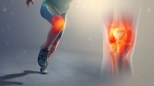
Knee is the most complex and the largest joint of the body. The joint consists of the upper end of the tibia (shinbone), lower end of the femur (thighbone), and the patella (kneecap). Ligaments and tendons bond the femur and the bones of the lower leg together. The four major ligaments in the knee connect the bones like strong ropes to hold them together.
The quadriceps tendons connect the muscles in the front of the thigh to the patella. Segments of the quadriceps tendon known as patellar retinacula are attached to the tibia that help to stabilize the patella. Articular cartilage, a slippery substance, covers the ends of the femur bone, the trochlear groove, and the underside of the patella.
The cartilage helps the bones to glide smoothly against each other for the movement of legs. Synovium which is a thin lining of tissue covering the surface of the joint produces a small amount of fluid helping in lubricating the cartilage. A small pad-like covering known as kneecap, acts like cushions and shock absorber.
Reasons causing patellofemoral pain syndrome are:
- Overuse of the knee joint is caused by strenuous physical activities which put stress on the knee like jogging, squatting, and climbing stairs
- Changes in kneecap alignment like shifting of the kneecap towards the outside or inside of the leg known as patella alta
- The quadriceps muscles along with quadriceps tendon keep the kneecap within the trochlear groove. Weak muscles surrounding the knee can lead to poor tracking of the kneecap within the groove.
- Improper equipment use while sports training
- Changes of the footwear or Excessively worn footwear
- The synovial irritation and inflammation causes changes in the distal femur or patella known as bone bruises
SYMPTOMS OF PATELLOFEMORAL PAIN SYNDROME :-
The syndrome is characterized as dull, aching pain in the front of the knee that begins gradually and is related to activities like climbing stairs, running, jumping, or squatting. Other symptoms along with pain are:
- Causes mild swelling
- Sensation like grating or grinding occurs when bending or extending the leg
- Thigh muscle strength is reduced of the initial symptoms are not treated on time
- Crackling or popping sounds while standing or climbing stairs
- Flexibility of the knee is reduced
The pain may be aggravated due to exercise and activities that repeatedly require to bend the knee.
DIAGNOSIS OF PATELLOFEMORAL PAIN SYNDROME
Physical examination like things that worsen the pain, check the kneecap for signs of instability and check the range-of-motion exercises etc. helps in diagnosing the syndrome. Doctors would recommend tests like imaging tests, such as X-rays and MRI scans to confirm the syndrome as well as rule out other medical conditions.
X-Ray – This technique visualizes the bone but it is less effective at screening soft tissues.
CT-Scan – This visualizes both bone as well as soft tissues. This delivers a much higher dose of radiation.
MRI – This provides detailed images of bones and soft tissues like the knee ligaments and cartilage to diagnose the damage to the knees.
TREATMENT OF PATELLOFEMORAL PAIN SYNDROME :-
The treatment aims to reduce the inflammation and pain to carry on the daily activities as well as sports training with ease and to increase the range of motion. Along with medications and therapies resting, avoiding or modifying the activities that increase the pain like climbing stairs, kneeling or squatting. The treatment regime is non-surgical or surgical depending on the extent of pain and severity of the condition.
Non-Surgical – This combines medications and therapies that reduces inflammation and reduces pain.
Medicines – Over-the-counter pain medication like acetaminophen (Tylenol, others), ibuprofen (Advil, Motrin IB, others) or naproxen sodium (Aleve) are prescribed to relieve pain and carry on daily activities. Non-steroidal anti-inflammatory drugs (NSAIDs) help in reducing pain in the short duration. Glycosaminoglycan polysulfate (GAGPS) inhibits proteolytic enzymes as well as increases the synthesis and range of polymerization of hyaluronic acid in synovial fluid which reduces pain and inflammation.
Physical Therapy – These exercises help improve the range of motion, strength, and endurance of the knee. The physiotherapists focus on strengthening and stretching the quadriceps muscles as these are main stabilizers of the kneecap. Core exercises help in strengthening the muscles of the abdomen and lower back.
Orthotics – The shoe inserts or insoles help to align and stabilize the foot and ankle relieving the stress off of your lower leg. These are available over-the-counter or can be custom made.
RICE (rest, ice, compression, and elevation) method – Wrapping the knee in an elastic bandage or using a pull-on bandage with the kneecap cut out helps reduce the inflammation and pain in the knee.
Taping or braces – taping or braces helps in reducing the pain and improves mechanism by mechanical realignment of the patella in the intertrochlear groove. This involves pulling patella medially with tape.
Surgical Treatment – This option for patellofemoral pain treatment is used very rarely and is done only for severe cases which do not respond to nonsurgical treatment. These may include:
Arthroscopy – This involves insertion of a pencil-thin device installed with a camera (known as arthroscope) into your knee through a tiny incision. This would remove the fragments of damaged cartilage.
Debridement – This would remove damaged articular cartilage from the surface of the patella, thereby providing relief from pain.
Lateral release – A lateral release procedure helps in pulling the patella out of the trochlear groove if the lateral retinaculum tendon is very tight. This can loosen the tissue thus correcting the patellar malalignment.
Tibial tubercle transfer – In some severe cases it is important to realign the kneecap which involves moving the patellar tendon along with a portion of the bony structure on the tibia (shinbone) known as tibial tubercle.
The patellofemoral pain is usually managed through simple measures. However, it can reoccur if proper care is not taken. Some precautions that avoid the recurrence are:
Shoes should be or proper fit and appropriate to your activities
Proper warm-up before physical activity that should include stretching and flexibility exercises for the quadriceps and hamstrings
- Appropriate stretching after physical activity
- Training should be increased gradually after recovery
- To be careful and avoid any activity that has damaged your knees in the past
OUTLOOK
The patellofemoral pain and dysfunction is often caused from either abnormal forces like increased pull of the lateral quadriceps retinaculum leading to subluxation or dislocation as well as repetitive compression or shearing forces like running or jumping. Surgical and non-surgical methods would ease the pain.
If you or anyone you know is suffering from patellofemoral pain syndrome, our expert providers at Specialty Care Clinics will take care of your health and help you recover.
Call us on 469-545-9983 to book an appointment with Dr. Raymond Fulp.