URETERAL OBSTRUCTION – SYMPTOMS, CAUSES, AND TREATMENT
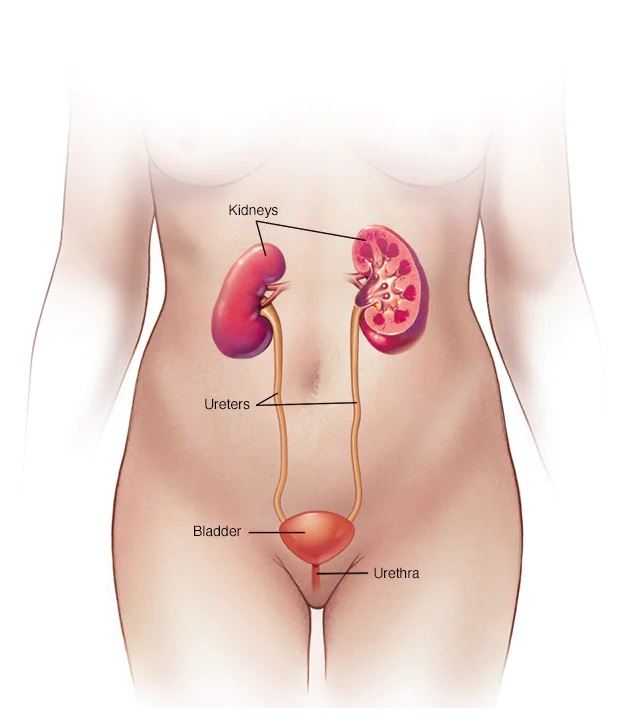
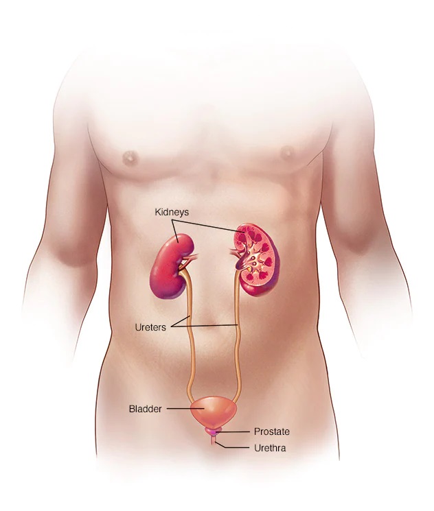
Ureteral obstruction is a block in one or both of the tubes (ureters) that carry urine from the kidneys to the bladder. The ureteral obstruction could be cured. But, if it is not treated, symptoms could quickly move from mild — pain, fever, and infection — to severe — loss of kidney function, sepsis, and death.
Ureteral obstruction is quite common. Because it is treatable, severe complications are uncommon.
SYMPTOMS
Ureteral obstruction may have no signs or symptoms. Signs and symptoms depend on where the obstruction happens, whether it is partial or complete, how quickly it develops, and whether it affects one or both kidneys.
Signs and symptoms may include:
- Pain
- Changes in the quantity of urine you produce (urine output)
- Difficulty urinating
- Blood in the urine
- Urinary tract infections
- High blood pressure (hypertension)
WHEN SHOULD YOU SEE A DOCTOR?
Schedule an appointment with your health care provider if you have signs and symptoms that worry you.
Look for medical attention if you experience:
- Pain so severe that you cannot sit still or find a comfortable place
- Pain followed by nausea and vomiting
- Pain followed by fever and chills
- Blood in your urine
- Difficulty passing urine
CAUSES
Different types of ureteral obstruction have different causes, some of which are present at birth (congenital). They include:
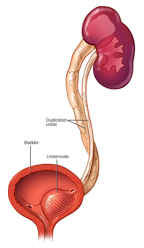
- A second (duplicated) ureter – This common condition, which is congenital, causes two ureters to develop on the same kidney. The second ureter could be completely or only partially developed. If either ureter does not work properly, urine could back up into the kidney and cause damage.
- A blockage (obstruction) where the ureter connects to the kidney or bladder – This stops urine flow. A blockage where the ureter and kidney meet (ureteropelvic junction) might cause the kidney to swell and ultimately stop working. This condition could be congenital or could develop with typical childhood growth, resulting from an injury or scarring, or in rare cases, develop from a tumor. A blockage where the ureter and bladder meet (ureterovesical junction) might cause urine to back up into the kidneys.
- Ureterocele – If a ureter is too narrow and does not allow urine to flow fully, a small bulge in the ureter (ureterocele) might develop. When a ureterocele develops, it is generally in the section of the ureter closest to the bladder. This could block urine flow and cause urine to back up into the kidney, possibly leading to kidney damage.
- Retroperitoneal fibrosis – This rare disorder happens when fibrous tissue grows in the area behind the abdomen. The fibers might grow as the result of cancer tumors or from taking specific medicines used to treat migraines. The fibers surround and block the ureters, causing urine to back up into the kidneys.
Other possible causes
Various causes inside (intrinsic) or outside (extrinsic) the ureter could lead to ureteral obstruction, including:
- Kidney stones
- Cancerous and non-cancerous tumors
- Blood clots
- Enlarged lymph nodes
- Internal tissue growth, like endometriosis in females
- Long-term swelling of the ureter wall, generally because of diseases like tuberculosis or a parasite infection known as schistosomiasis
COMPLICATIONS
The ureteral obstruction could lead to urinary tract infections and kidney damage, which could be irreversible.
DIAGNOSIS
Generally, providers diagnose ureteral obstruction disorders before birth during routine prenatal ultrasounds, which could show details of the developing fetus, including the kidneys, ureters, and bladder. Providers usually perform another ultrasound after birth to re-evaluate the kidneys.
If your provider suspects you have an obstructed ureter, some of these tests and scans may be used to reach a diagnosis:
- Blood and urine tests – Your provider checks samples of your blood and urine for signs of infection and the presence of creatinine, which signals that your kidneys are not working properly.
- Ultrasound – An ultrasound of the area behind your abdominal organs (retroperitoneal ultrasound) enables your provider to view the kidneys and ureters.
- Bladder catheterization – To test for incomplete or blocked urine flow, your provider inserts a tiny tube (catheter) through the urethra, administers dye into your bladder, and takes X-rays of your kidneys, ureters, bladder, and urethra prior to and during urination.
- Renal nuclear scan – Your provider or a technician administers a tracer that contains a tiny amount of radioactive material into your arm. A special camera detects radioactivity and produces pictures that your provider uses to evaluate the urinary system.
- Cystoscopy – A tiny tube with a camera and light is inserted into your urethra or through a tiny incision. The optical system enables the provider to see inside the urethra and bladder.
- Computerized tomography (CT) scan – A CT scan combines a series of X-ray views taken from many different angles and computer processing to create cross-sectional pictures of your kidneys, ureter, and bladder.
- Magnetic resonance imaging (MRI) – An abdominal MRI uses a magnetic field and radio waves to create detailed pictures of the organs and tissues that make up your urinary system.
TREATMENT
The aim of ureteral obstruction treatment is to remove blockages, if possible, or bypass the blockage, which might help repair damage to the kidneys. Treatment may include antibiotics to clear related infections.
Drainage procedures
A ureteral obstruction that causes severe pain may need an immediate procedure to remove urine from your body and temporarily ease the problems caused by a blockage. Your doctor (urologist) might recommend:
- A ureteral stent is a hollow tube inserted within the ureter to keep it open.
- Percutaneous nephrostomy, in which your doctor inserts a tube through your back to drain the kidney directly (percutaneous nephrostomy).
- A catheter is a tube inserted through the urethra to join the bladder to an external drainage bag. This might be particularly important if problems with your bladder also contribute to poor drainage of your kidneys.
Your doctor could tell you which procedure or combination of procedures is best for you. Drainage procedures may provide temporary or permanent relief, depending on your condition.
Surgical procedures
There are a lot of surgical procedures used to correct ureteral obstructions. The kind of procedure depends on your situation.
Ureteral obstruction surgery might be performed through one of these surgical approaches:
- Endoscopic surgery – This minimally invasive procedure includes passing a lighted scope through the urethra into the bladder and other parts of the urinary tract. The surgeon makes a cut into the affected or blocked part of the ureter to expand the area and then places a hollow tube (stent) in the ureter to keep it open. This procedure might be performed to both diagnose and treat a condition.
- Open surgery – The surgeon makes a cut in your abdomen to remove the blockage and repair your ureter.
- Laparoscopic surgery – In this approach, the surgeon makes one or more tiny incisions through your skin to insert a tiny tube with a light, a camera, and other instruments required for the procedure.
- Robot-assisted laparoscopic surgery – The surgeon employs a robotic system to perform a laparoscopic procedure.
The primary differences among these surgical approaches are your recovery time after surgery and the number and size of incisions used for the procedure. Your doctor (urologist) determines the type of procedure and the best surgical approach for treating your condition.
If you or anyone you know is suffering from ureteral obstruction, our expert providers at Specialty Care Clinics will take care of your health and help you recover.
Call us on (469) 545-9983 to book an appointment with our specialists.