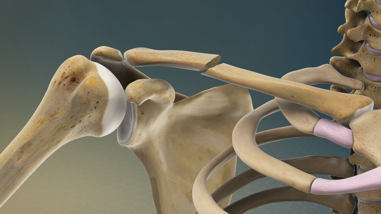
Understanding the Clavicle: Types of Fractures and Treatment Options
The clavicle, commonly known as the collarbone, is a crucial bone in the human body that plays a significant role in supporting the shoulder and facilitating a wide range of arm movements. Unfortunately, like any other bone, the clavicle is susceptible to fractures. In this comprehensive article, we will explore the anatomy of the clavicle, delve into the various types of clavicle fractures, and discuss the treatment options available for individuals who experience this injury.
Anatomy of the Clavicle:
Structure and Function
The clavicle is an S-shaped bone that connects the sternum (breastbone) to the scapula (shoulder blade). Its distinctive shape provides structural support to the shoulder, allowing for optimal movement and functionality of the upper limb. The clavicle acts as a strut, holding the shoulder in place and facilitating the transmission of forces from the arm to the axial skeleton.
Joints and Ligaments
Articulating with the sternum at the sternoclavicular joint and the scapula at the acromioclavicular joint, the clavicle forms critical connections in the shoulder girdle. Ligaments surrounding these joints contribute to stability, ensuring the coordinated movement of the upper extremity.
Blood Supply and Nerve Innervation
Blood supply to the clavicle comes primarily from branches of the subclavian artery. Nerves, including branches of the brachial plexus, innervate the clavicle, contributing to its sensory and motor functions.
Types of Clavicle Fractures:
Midshaft Fractures
Midshaft fractures are the most common type of clavicle fractures, accounting for approximately 80% of cases. These fractures typically occur due to a direct blow to the shoulder or a fall onto an outstretched hand. Symptoms include pain, swelling, and bruising over the fracture site.
Proximal Fractures
Proximal clavicle fractures involve the region near the sternoclavicular joint. These fractures may result from high-energy trauma, such as a motor vehicle accident, or a fall onto the shoulder. Displacement of the fracture may affect nearby structures, including blood vessels and nerves.
Distal Fractures
Distal clavicle fractures occur near the acromioclavicular joint and are less common than midshaft fractures. These fractures are often associated with trauma or a fall onto the outstretched hand, leading to pain and limited shoulder movement.
Greenstick Fractures
Greenstick fractures are incomplete fractures where the bone bends but does not break completely. While more common in children due to their softer bones, adults can also experience greenstick fractures, especially in the clavicle.
Diagnosis and Imaging
Clinical Examination
Diagnosing a clavicle fracture begins with a thorough clinical examination, including assessing the range of motion, tenderness, and deformity at the fracture site. Physicians may also evaluate neurological and vascular function to identify any associated injuries.
Radiographic Imaging
X-rays are the primary diagnostic tool for clavicle fractures. Anteroposterior and lateral views help determine the location, displacement, and alignment of the fracture. Advanced imaging modalities such as CT scans or MRI may be used for more detailed assessments.
Treatment Options
Conservative Management
Many clavicle fractures can be treated conservatively, especially if the bones are well-aligned. Immobilization with a sling or figure-eight brace helps reduce movement, allowing the bones to heal naturally. Physical therapy may be recommended to restore strength and range of motion.
Surgical Intervention
Surgery may be considered for displaced or complex fractures. Common surgical procedures include open reduction and internal fixation (ORIF) using plates, screws, or pins to stabilize the fractured bones. Surgical intervention is often preferred for high-energy injuries or fractures with significant displacement.
Rehabilitation and Follow-up
Rehabilitation plays a crucial role in the recovery process. Physical therapy helps restore shoulder function, strengthen muscles, and prevent stiffness. Regular follow-up appointments and imaging assessments monitor the progress of healing and ensure that any complications are promptly addressed.
Conclusion
In conclusion, understanding the clavicle’s anatomy, recognizing the types of fractures that can occur, and being aware of the available treatment options are essential for both medical professionals and the general public. Clavicle fractures, while common, can have varying degrees of severity, necessitating tailored approaches to management. By staying informed about the intricacies of clavicle fractures, individuals can make informed decisions regarding their health and well-being. Whether treated conservatively or through surgical intervention, the goal is to promote optimal recovery and restore the clavicle’s vital role in supporting the intricate mechanics of the shoulder.


